What do we know?
Skin is a special organ that both protects us and allows us to sense the world around us.
Skin is made of three layers, each containing specialised cells.
A variety of stem cells are needed to maintain and repair our skin on a daily basis. Researchers have identified stem cells responsible for making the epidermal layer, hair follicles and skin pigments.
Epidermal stem cells are currently used in clinics to grow the outer layer of skin (epidermis) for patients with life-threatening burns and genetic disorders. However, the process is difficult and expensive. Moreover, if the skin is severely damaged, for example by burns, the transplanted skin will lack sweat glands, hair follicles and sebaceous (oil) glands.
What are researchers working on?
Researchers are currently working to develop methods to grow skin that contains more of the normal functional components, such as sebaceous glands and hair follicles. This will make skin grafts more durable and natural-looking.
Currently, lab-grown epidermis typically relies on animal cells as a support for the growth of human skin epidermal cells. While this approach has been shown to be safe, researchers are actively working to develop alternatives that do not require animal cells for treatments.
Researchers are also working on using genetically modified skin stem cells to treat skin diseases, such as epidermolysis bullosa.
What are the challenges?
Recently, great progress has been made on growing skin that contains components such as hair follicles and glands. However, our body has many different types of skin; just compare the palms of your hand to your scalp. Learning how to grow these different types of skin will be an important challenge to overcome.
The largest challenge for developing skin stem cell treatments is creating methods that are readily available and affordable for patients.
The structure of skin
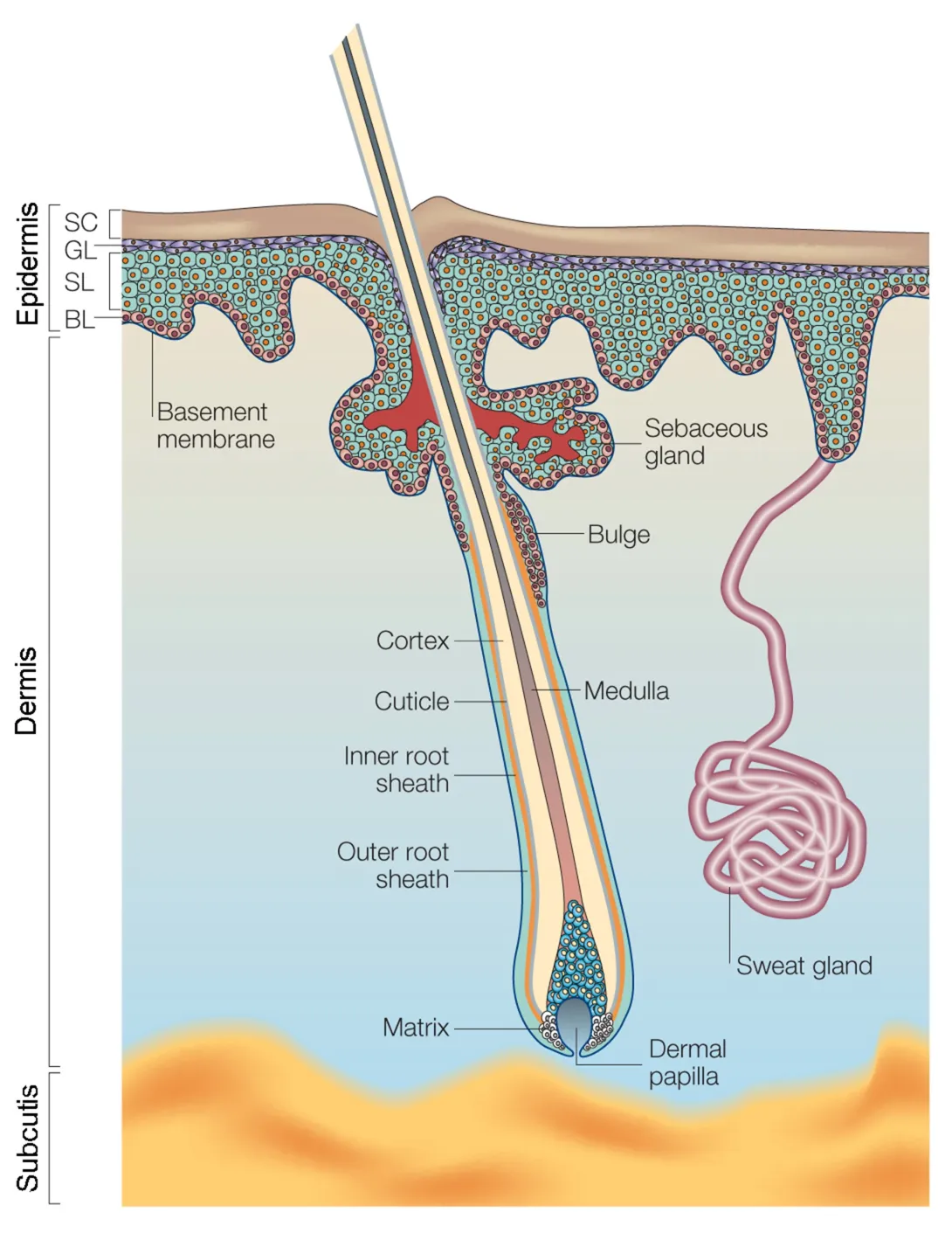
In humans and other mammals, the skin has three parts - the epidermis, the dermis and the subcutis (or hypodermis). The epidermis forms the surface of the skin. It is made up of several layers of cells called keratinocytes. The dermis lies underneath the epidermis and contains skin appendages: hair follicles, sebaceous (oil) glands and sweat glands. The subcutis contains fat cells and some sweat glands.

Layers of the skin
The skin has three main layers - the epidermis, dermis and subcutis. The epidermis contains layers of cells called keratinocytes. BL = basal layer; SL = spinous layer; GL = granular layer; SC= stratum corneum.
In everyday life your skin has to cope with a lot of wear and tear. For example, it is exposed to chemicals like soap and to physical stresses such as friction from your clothes or exposure to sunlight. The epidermis and skin appendages need to be renewed constantly to keep your skin in good condition. What’s more, if you cut or damage your skin, it has to be able to repair itself efficiently to keep doing its job – protecting your body from the outside world.
Skin stem cells make all this possible. They are responsible for constant renewal (regeneration) of your skin, and for healing wounds. So far, scientists have identified several different types of skin stem cell:
- Epidermal stem cells are responsible for everyday regeneration of the different layers of the epidermis. These stem cells are found in the basal layer of the epidermis.
- Hair follicle stem cells ensure constant renewal of the hair follicles. They can also regenerate the epidermis and sebaceous glands if these tissues are damaged. Hair follicle stem cells are found throughout the hair follicles.
- Melanocyte stem cells are responsible for regeneration of melanocytes, a type of pigment cell. Melanocytes produce the pigment melanin, and therefore play an important role in skin and hair follicle pigmentation. It is not yet certain where these stem cells are found in humans.
Some studies have also suggested that the dermis and hypodermis contain stem cells known as MSCs. This remains controversial among scientists, and further studies are needed to determine whether these cells are truly stem cells and what their role is in the skin.
Epidermal stem cells are one of the few types of stem cell already used to treat patients. Thanks to a discovery made in 1970 by Professor Howard Green in the USA, epidermal stem cells can be taken from a patient, multiplied and used to grow sheets of epidermis in the lab. The new epidermis can then be transplanted back onto the patient as a skin graft. This technique is mainly used to save the lives of patients who have third degree burns over very large areas of their bodies. Only a few clinical centres are able to carry out the treatment successfully, and it is an expensive process. It is also not a perfect solution. Only the epidermis can be replaced with this method; the new skin has no hair follicles, sweat glands or sebaceous glands.
One of the current challenges for stem cell researchers is to understand how all the skin appendages are regenerated. This could lead to improved treatments for burn patients, or others with severe skin damage.
Researchers are working to identify new ways to grow skin cells in the lab. Epidermal stem cells are currently cultivated on a layer of fibroblast cells from rodents, called feeder cells. These cell culture conditions have been proved safe, but it would be preferable to avoid using animal products when cultivating cells that will be transplanted into patients. So, researchers are searching for effective cell culture conditions that will not require the use of rodent cells.
Scientists are also working to treat genetic diseases affecting the skin. Since skin stem cells can be cultivated in laboratories, researchers can genetically modify the cells, for example by inserting a working copy of a mutated gene. The correctly modified cells can be selected, grown and multiplied in the lab, then transplanted back onto the patient. Epidermolysis Bullosa is one example of a genetic skin disease where patients can benefit from this approach; work is underway to test the technique.
